
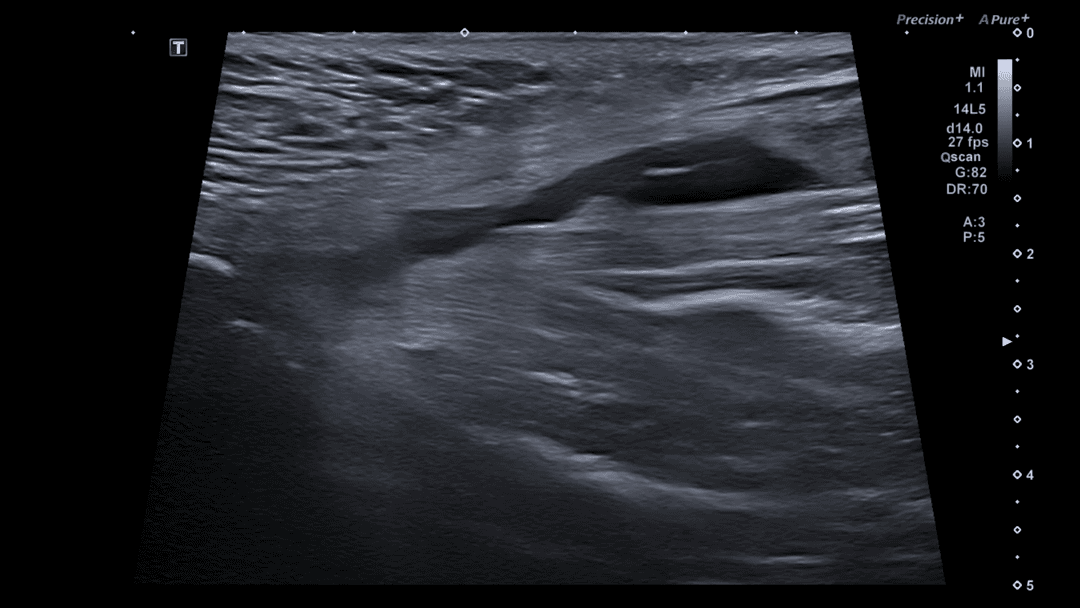
Adductor Longus Muscle Tear
Last updated
Learn this and more in our online course: Point-of-Care Ultrasound for Pain Diagnosis: Hip to Knee
Typical Mechanism: Often results from acute overstretching during forceful hip abduction, especially common in sports involving rapid directional changes (soccer, hockey).
Sonographic Appearance: Look for disruption of muscle fibers, hematoma formation, hypoechoic gaps, and loss of the normal pennate muscle pattern. Fluid collections may be evident between disrupted fibers.
Key Anatomical Landmark: Evaluate carefully at the musculotendinous junction, located near the pubic attachment; tears frequently occur proximally.
Dynamic Assessment: Utilize gentle active and passive hip abduction and adduction during ultrasound to confirm tear presence, assess fiber discontinuity, and distinguish partial from complete tears.
Want to deepen your ultrasound skills?
Explore our hands-on and online courses in musculoskeletal ultrasound.