
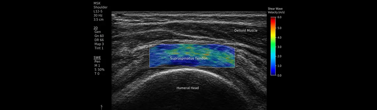
The Next Dimension in Tendon Imaging: Shear Wave Elastography in Musculoskeletal Ultrasound
Discover how Shear Wave Elastography (SWE) is transforming the assessment of tendons in musculoskeletal ultrasound. This evidence-based webinar combines current research with practical clinical application to help clinicians confidently interpret tendon stiffness and integrate SWE into everyday practice.
What you'll learn
- Principles of Shear Wave Elastography
- Optimising SWE scanning techniques
- Normal vs pathological tendon stiffness (B-mode and SWE)
- B-mode and SWE in tendinopathy and post-operative tendon healing
- Clinical interpretation and common pitfalls
- Real case studies and practical MSK applications
Meet Your Trainers

Dates & prices
- 13 April '2719:00 – 20:3060-days access to the recording€ 127,50
- 13 April '2719:00 – 20:3030-days access to the recording€ 97,50
About this webinar
Shear Wave Elastography (SWE) is new, exciting, and challenging the way we have traditionally assessed tendons. For more than 15 years, my research with SWE has explored what lies beyond conventional ultrasound morphology. While B-mode ultrasound shows us structure, SWE adds another dimension: tissue stiffness. It allows us to move beyond simply looking at a tendon and begin to understand what is happening within it.
In this webinar, we will challenge you to explore the “real” tendon and discover how SWE can add meaningful clinical information to the assessment of tendon pathology and healing. Turn black and white into colour, see beyond the image, and measure what you cannot see.